Kidney stone disease
Kidney stone disease, also known as renal calculus disease, nephrolithiasis, or urolithiasis, involves the formation of solid mineral 'stones' within the urinary tract. These stones typically form in the kidneys and can pass through the urinary tract. Small stones often pass without symptoms, but larger stones can cause severe pain, blood in the urine, vomiting, or painful urination.
Signs and Symptoms
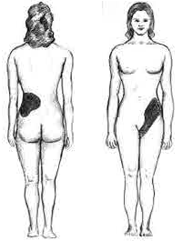
The hallmark of kidney stones is excruciating, intermittent pain radiating from the flank to the groyne or inner thigh, known as renal colic. Pain is typically accompanied by urinary urgency, restlessness, hematuria (blood in the urine), sweating, nausea, and vomiting. The pain comes in waves lasting 20 to 60 minutes due to peristaltic contractions of the ureter as it attempts to expel the stone. Pain in the lower-left quadrant can sometimes be confused with diverticulitis.
Risk Factors
Dehydration, high dietary intake of animal protein, sodium, sugars, and certain metabolic conditions like hyperparathyroidism and gout can increase the risk of stone formation. Obesity, sedentary lifestyles, and living in warm climates also contribute to higher risk. Some medications, such as calcium supplements, and conditions like Crohn's disease are associated with kidney stones.

Diagnosis
Diagnosis is based on symptoms, urine testing, and medical imaging. Imaging studies include noncontrast helical CT scans, which are the diagnostic method of choice. Renal ultrasonography can also be useful, especially in children or pregnant women.

Laboratory investigations include microscopic examination of urine, urine culture, complete blood count, renal function tests, and 24-hour urine collection. Chemical analysis of stones can guide preventive and therapeutic management.

Prevention
Preventive measures depend on the type of stones. General recommendations include increasing fluid intake to produce more than two litres of urine per day and avoiding soft drinks containing phosphoric acid. Specific dietary adjustments and medications like thiazide diuretics, citrate, or allopurinol may also be recommended.
Dietary Measures
Preventive strategies involve a combination of dietary modifications and medications. Recommendations include:
- Increasing fluid intake to achieve more than two litres per day of urine output.
- Limiting animal protein intake.
- Increasing citrate intake from sources like lemon and lime juice.
- Reducing sodium intake.
Urine Alkalinisation
For uric acid stones, alkalinisation of urine using medications or dietary supplements like alkali citrate, sodium bicarbonate, or potassium citrate is recommended. Testing urine pH periodically can help maintain optimal conditions for stone dissolution.
Treatment
Pain Management
Pain management often requires intravenous NSAIDs or opioids. Oral medications can be effective for less severe pain, and the use of antispasmodics does not provide additional benefits.
Medical Expulsive Therapy
Medications like alpha adrenergic blockers (e.g., tamsulosin) or calcium channel blockers (e.g., nifedipine) can speed the spontaneous passage of stones in the ureter. These medications may reduce pain, hospital visits, and the need for further interventions.
Lithotripsy
Extracorporeal shock wave lithotripsy (ESWL) is a noninvasive technique for breaking kidney stones using high-intensity pulses of ultrasonic energy. It is effective for stones near the renal pelvis and upper ureter, provided the stone burden is less than 20 mm.

Surgery
For stones that do not pass spontaneously or cause severe complications, surgical interventions may be necessary. Techniques include ureteroscopy, which involves the use of a stent to bypass an obstruction, and more invasive procedures like percutaneous nephrolithotomy for large or complicated stones.

Kidney stone disease is a common and recurring condition that requires careful management and preventive strategies. Understanding its risk factors, diagnostic methods, and treatment options is very important for effective patient care.
Self-assessment MCQs (single best answer)
What is the hallmark symptom of kidney stones?

Which imaging study is the diagnostic method of choice for kidney stones?
Which dietary measure is recommended to prevent kidney stones?
Which medication can be used for the alkalinisation of urine to prevent uric acid stones?
What is the purpose of extracorporeal shock wave lithotripsy (ESWL)?
Which of the following is NOT a risk factor for kidney stone formation?
Which laboratory investigation is used for guiding preventive and therapeutic management of kidney stones?
What type of crystals might be found on microscopic examination of the urine in patients with kidney stones?
Which medication can be used to facilitate the spontaneous passage of stones in the ureter?
For which condition might a ureteral stent be used as a treatment?
Dentaljuce
Dentaljuce provides Enhanced Continuing Professional Development (CPD) with GDC-approved Certificates for dental professionals worldwide.
Founded in 2009 by the award-winning Masters team from the School of Dentistry at the University of Birmingham, Dentaljuce has established itself as the leading platform for online CPD.
With over 100 high-quality online courses available for a single annual membership fee, Dentaljuce offers comprehensive e-learning designed for busy dental professionals.
The courses cover a complete range of topics, from clinical skills to patient communication, and are suitable for dentists, nurses, hygienists, therapists, students, and practice managers.
Dentaljuce features Dr. Aiden, a dentally trained AI-powered personal tutor available 24/7 to assist with queries and provide guidance through complex topics, enhancing the learning experience.
Check out our range of courses, or sign up now!