Osteosarcoma
Osteosarcoma (OS), also known as osteogenic sarcoma (OGS), is a cancerous tumour in bone characterised by aggressive malignant neoplasm originating from primitive transformed mesenchymal cells. These cells exhibit osteoblastic differentiation and produce malignant osteoid. Osteosarcoma is the most common histological form of primary bone sarcoma, predominantly affecting teenagers and young adults.
Signs and Symptoms
Patients often first complain of pain, which may be worse at night, intermittent, and of varying intensity, and may have persisted for a long time. Active teenagers frequently report pain in the lower femur or just below the knee. Large tumours can present as overt localised swelling, and sudden fractures can occur due to weakened bones. Deep-seated tumours, such as those in the pelvis, may not show apparent swelling.

Causes
Research indicates potential roles for cancer stem cells, specific genes, and proteins in tumour development. Familial genetic factors such as deletion of chromosome 13q14 and conditions like Li-Fraumeni syndrome and Rothmund-Thomson syndrome are associated with higher risks. Environmental factors, such as large doses of Sr-90, might also contribute. However, there is no clear association between water fluoridation and osteosarcoma.
Mechanism
Osteosarcomas typically occur at bone growth sites, possibly due to the susceptibility of proliferative osteoblastic cells to mutations. Commonly affected areas include the proximal tibia, humerus, and distal femur, with the tumour exhibiting a solid, hard, irregular appearance on X-rays, often described as "sun-burst" or "moth-eaten."

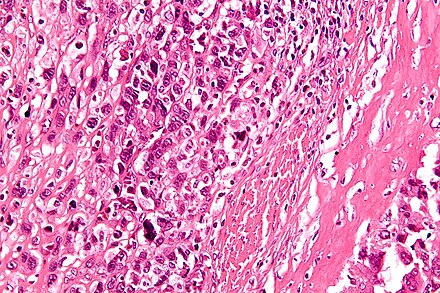
Microscopically, osteosarcoma is characterised by the presence of osteoid within the tumour, with tumour cells being highly pleomorphic and producing irregular trabeculae.
Diagnosis
Diagnosis begins with X-rays, revealing characteristic sunburst appearances and Codman triangles. CT scans help define the bony anatomy and detect pathological fractures, while MRI scans are better for imaging soft tissues and medullary cavities. Dentists may first detect osteosarcoma during routine check-ups, particularly in the mandible, and refer patients for further evaluation and biopsy.

Treatment
The primary treatment for osteosarcoma is complete radical surgical resection. Neoadjuvant chemotherapy is typically administered before surgery, followed by postoperative chemotherapy. Common chemotherapy agents include high-dose methotrexate, cisplatin, adriamycin, and ifosfamide. Mifamurtide may be used post-surgery to reduce recurrence risk. Limb-salvage surgery is preferred, but complications might necessitate amputation.
Prognosis
Prognosis varies by stage. Stage I osteosarcoma has an excellent prognosis with wide resection. Stage II prognosis depends on tumour site, size, necrosis degree, and other pathological factors. Stage III prognosis hinges on the resectability of primary tumours and lung metastases. Survival rates have improved significantly, with long-term survival probabilities around 68%.
Epidemiology
Osteosarcoma is the eighth-most common childhood cancer, comprising 2.4% of paediatric malignancies and 20% of primary bone cancers. Incidence rates in the U.S. are estimated at 5.0 per million per year for individuals under 20, with a slightly higher prevalence in males and certain ethnic groups.
Other Animals
Osteosarcoma is also prevalent in dogs, particularly large breeds, and affects similar bone sites as in humans. Treatment often involves amputation and chemotherapy, but survival time remains limited. In cats, the disease is less aggressive, and amputation alone can result in significant survival time.

Self-assessment MCQs (single best answer)
Which age group is predominantly affected by osteosarcoma?

Which of the following is NOT a common sign or symptom of osteosarcoma?
What are the primitive transformed cells in osteosarcoma known for producing?
Which genetic condition is associated with a higher risk of developing osteosarcoma?
On an X-ray, osteosarcoma often presents with which characteristic appearance?
Which imaging modality is preferred for detailed imaging of soft tissues and medullary cavities in osteosarcoma?
What is the primary treatment approach for osteosarcoma?
Which chemotherapy agent is commonly used in the treatment of osteosarcoma?
What is the typical prognosis for stage I osteosarcoma with wide resection?
In which animal is osteosarcoma most prevalent, particularly affecting similar bone sites as in humans?
Dentaljuce
Dentaljuce provides Enhanced Continuing Professional Development (CPD) with GDC-approved Certificates for dental professionals worldwide.
Founded in 2009 by the award-winning Masters team from the School of Dentistry at the University of Birmingham, Dentaljuce has established itself as the leading platform for online CPD.
With over 100 high-quality online courses available for a single annual membership fee, Dentaljuce offers comprehensive e-learning designed for busy dental professionals.
The courses cover a complete range of topics, from clinical skills to patient communication, and are suitable for dentists, nurses, hygienists, therapists, students, and practice managers.
Dentaljuce features Dr. Aiden, a dentally trained AI-powered personal tutor available 24/7 to assist with queries and provide guidance through complex topics, enhancing the learning experience.
Check out our range of courses, or sign up now!