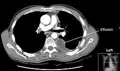
Pleural Effusion
Pleural effusion is the accumulation of excess fluid in the pleural cavity, the potential space that surrounds each lung.
Normally, pleural fluid is secreted by the parietal pleural capillaries and cleared by lymphatic absorption, leaving only 5–15 millilitres of fluid to maintain a functional vacuum between the parietal and visceral pleurae.
Excess fluid can impair inspiration by disrupting this vacuum, increasing resistance against lung expansion, and potentially causing partial or complete lung collapse.

Types and Causes
Pleural effusions can be classified based on the type of fluid or the underlying cause. Types of fluid include serous fluid (hydrothorax), blood (hemothorax), pus (pyothorax or empyema), chyle (chylothorax), and more rarely, urine (urinothorax). By pathophysiology, pleural effusions are categorised as transudative or exudative.
Transudative Pleural Effusion

Transudative effusions are often caused by systemic factors such as increased hydrostatic pressure or decreased colloid osmotic pressure. Common causes include congestive heart failure, liver cirrhosis, nephrotic syndrome, and severe hypoalbuminaemia. Conditions like acute atelectasis, myxedema, peritoneal dialysis, and end-stage kidney disease can also lead to transudative effusions.
Exudative Pleural Effusion

Exudative effusions are caused by local factors such as inflammation, infection, or malignancy, leading to increased capillary permeability. Common causes include bacterial pneumonia, cancer (notably lung cancer, breast cancer, and lymphoma), viral infections, and pulmonary embolism. Other causes include trauma, autoimmune disorders, pancreatitis, and ruptured oesophagus.
Signs and Symptoms
A pleural effusion can present with symptoms such as dyspnea, chest pain, and cough. Physical examination may reveal decreased movement of the chest on the affected side, dullness to percussion, diminished breath sounds, decreased vocal resonance, and pleural friction rub.
Diagnosis

Diagnosis typically involves medical history review, physical examination, and imaging studies like chest X-rays and computed tomography (CT). Ultrasound is increasingly used for its accuracy and ability to provide dynamic imaging. Chest radiographs in the lateral decubitus position can detect smaller effusions than upright X-rays.
Imaging


Thoracentesis
Thoracentesis involves the insertion of a needle to draw pleural fluid for analysis. This procedure helps determine the cause of the effusion and involves tests such as chemical composition, Gram stain and culture, cell counts, and cytopathology.
Light's Criteria
Transudate vs. Exudate
| Criteria | Transudate | Exudate |
|---|---|---|
| Main Causes | ↑ hydrostatic pressure, ↓ colloid osmotic pressure | Inflammation-Increased vascular permeability |
| Appearance | Clear | Cloudy |
| Specific gravity | < 1.012 | > 1.020 |
| Protein content | < 2.5 g/dL | > 2.9 g/dL |
| Fluid protein/serum protein | < 0.5 | > 0.5 |
| SAAG = Serum [albumin] - Effusion [albumin] | > 1.2 g/dL | < 1.2 g/dL |
| Fluid LDH upper limit for serum | < 0.6 or < 2/3 | > 0.6 or > 2/3 |
| Cholesterol content | < 45 mg/dL | > 45 mg/dL |
| Radiodensity on CT scan | 2 to 15 HU | 4 to 33 HU |
Light's criteria help differentiate between transudative and exudative pleural effusions based on the chemical analysis of pleural fluid compared to blood.
Treatment
Treatment depends on the underlying cause. Therapeutic aspiration may suffice for smaller effusions, while larger ones may require an intercostal drain. Recurrent effusions might necessitate pleurodesis, a procedure to scar the pleural surfaces together, preventing fluid accumulation. In cases of malignant pleural effusion, indwelling pleural catheters like the PleurX Pleural Catheter can be used for outpatient fluid management. Tubercular pleural effusions are treated with antituberculosis therapy following specific regimens.

Self-assessment MCQs (single best answer)
Which of the following types of fluid is NOT typically associated with pleural effusion?
What is the most common cause of transudative pleural effusion?
Which of the following symptoms is NOT commonly associated with pleural effusion?
What is the primary purpose of thoracentesis in the context of pleural effusion?
Which imaging modality is increasingly used for its accuracy and dynamic imaging capability in diagnosing pleural effusion?
According to Light's criteria, which of the following is a characteristic of an exudative pleural effusion?
Which of the following is NOT a typical cause of exudative pleural effusion?
What treatment might be necessary for recurrent pleural effusions?
Which protein content level in pleural fluid suggests a transudative pleural effusion according to Light's criteria?
Which condition could lead to a transudative pleural effusion?
Dentaljuce
Dentaljuce provides Enhanced Continuing Professional Development (CPD) with GDC-approved Certificates for dental professionals worldwide.
Founded in 2009 by the award-winning Masters team from the School of Dentistry at the University of Birmingham, Dentaljuce has established itself as the leading platform for online CPD.
With over 100 high-quality online courses available for a single annual membership fee, Dentaljuce offers comprehensive e-learning designed for busy dental professionals.
The courses cover a complete range of topics, from clinical skills to patient communication, and are suitable for dentists, nurses, hygienists, therapists, students, and practice managers.
Dentaljuce features Dr. Aiden, a dentally trained AI-powered personal tutor available 24/7 to assist with queries and provide guidance through complex topics, enhancing the learning experience.
Check out our range of courses, or sign up now!


