Enhanced Verifiable CPD from the
University of Birmingham

Histology
Adrenal gland

Many of the slides below are optimised for colour-blind people - click the spectrum icon.

Click here to scroll down to the accompanying text and MCQs.

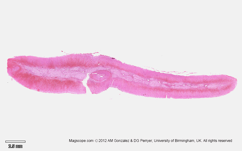
Micrograph of a cross section of the human adrenal illustrating the three cortical zones and the medulla.
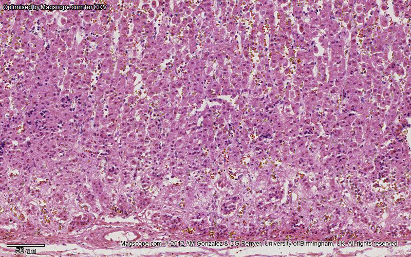
Light micrograph of the adrenal cortex illustrating the capsule, zona glomerulosa and zona fasciculata
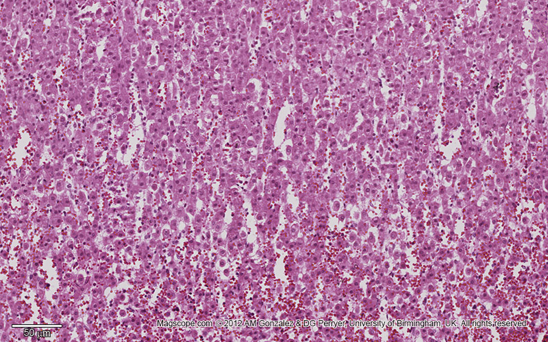
Light micrograph of the adrenal cortex illustrating the zona fasciculata
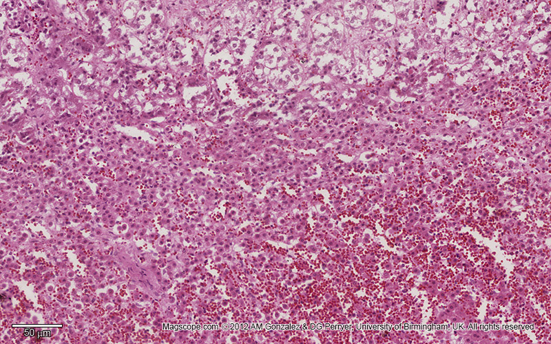
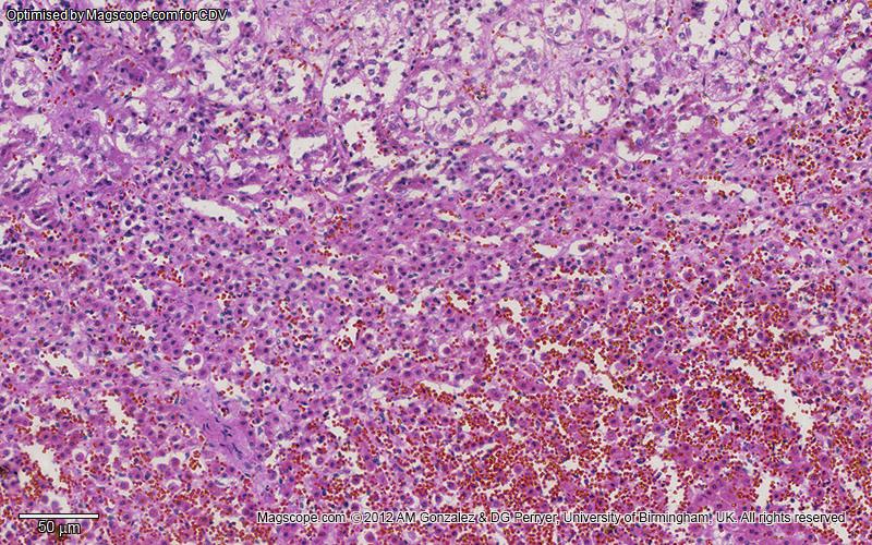
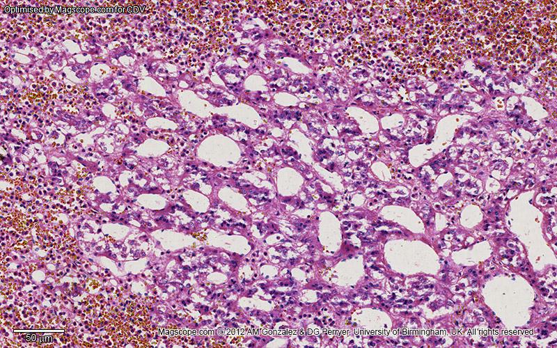
Light micrograph of the adrenal cortex illustrating the zona reticularis
Light micrograph illustrating the adrenal medulla

Adrenal Gland Histology and Histopathology

The adrenal glands, also known as suprarenal glands, are very important endocrine organs located above the kidneys. Each gland consists of two main parts: the outer adrenal cortex and the inner adrenal medulla, both producing essential hormones.

Position of adrenal gland

Adrenal Cortex

Zona Glomerulosa

The outermost layer of the adrenal cortex is the zona glomerulosa. Histologically, this layer features cells arranged in oval clusters or arcs and is responsible for producing mineralocorticoids such as aldosterone. The cells in this layer contain:

  • Oval groups of cells separated by strands of connective tissue.
  • Wide capillaries for efficient hormone transport.

Zona Fasciculata

Situated between the zona glomerulosa and zona reticularis, the zona fasciculata is the largest layer, constituting approximately 80% of the cortex's volume. The cells here are arranged in radially oriented columns and are packed with lipid droplets, mitochondria, and smooth endoplasmic reticulum, reflecting their role in producing glucocorticoids such as cortisol. The cells are:

  • Columnar in arrangement.
  • Rich in lipid droplets.
  • Abundant in mitochondria and smooth ER.

Zona Reticularis

The innermost layer, the zona reticularis, lies adjacent to the medulla and produces androgens. The cells in this layer form irregular cords and clusters, separated by capillaries and connective tissue. They contain:

  • Small amounts of cytoplasm.
  • Few lipid droplets.
  • Brown lipofuscin pigment at times.
Section of human adrenal gland under the microscope
Section of human adrenal gland under the microscope, showing its different layers. From the surface to the centre: zona glomerulosa, zona fasciculata, zona reticularis, medulla.

Adrenal Medulla

The adrenal medulla is located at the centre of each adrenal gland and is composed of chromaffin cells, which are the primary source of catecholamines such as adrenaline and noradrenaline. These cells are characterised by:

  • Dense network of blood vessels for rapid hormone release.
  • Chromaffin granules that stain with chromium salts.
  • Specialised sympathetic ganglion cells that lack distinct synapses and release secretions directly into the blood.

Blood Supply and Variability

The adrenal glands have an extensive blood supply, with up to 60 small arteries entering each gland. These include:

  • Superior suprarenal artery from the inferior phrenic artery.
  • Middle suprarenal artery from the abdominal aorta.
  • Inferior suprarenal artery from the renal artery.

Venous drainage is through the suprarenal veins:

  • Right suprarenal vein draining into the inferior vena cava.
  • Left suprarenal vein draining into the left renal vein or left inferior phrenic vein.

Histopathology

Corticosteroid Overproduction

Cushing's Syndrome

Cushing's syndrome results from glucocorticoid excess. Histologically, it can reveal increased cellularity and enlarged adrenal glands. Causes include ACTH-secreting pituitary adenomas and adrenal hyperplasia.

Primary Aldosteronism

This condition involves overproduction of aldosterone due to bilateral hyperplasia or adenomas. Histological examination shows hyperplastic zona glomerulosa or aldosterone-producing adenomas.

Adrenal Insufficiency

Addison's Disease

Addison's disease is characterised by adrenal cortex destruction, often appearing histologically as lymphocytic infiltration and fibrous tissue. Autoimmune reactions or infections like tuberculosis are common causes.

Characteristic skin hyperpigmentation in Addison's disease

Adrenal Tumours

Adrenal tumours, often incidental findings, include benign adenomas and rare malignant carcinomas. Histopathologically, adenomas are well-circumscribed, while carcinomas display cellular atypia and invasion.

Incidences and prognoses of adrenal tumours
Incidences and prognoses of adrenal tumours.


Self-assessment MCQs (single best answer)

Which layer of the adrenal cortex is responsible for producing mineralocorticoids such as aldosterone?


What is the primary hormone produced by the adrenal medulla?


Histologically, the cells of the zona fasciculata are characterised by:


Addison's disease is characterised by destruction of which part of the adrenal gland?


Which artery does NOT supply blood to the adrenal glands?


Which of the following is a feature of primary aldosteronism?


Chromaffin cells are primarily found in which part of the adrenal gland?


What is a common cause of Cushing's syndrome?


Which venous structure drains the right adrenal gland?


Which feature is commonly observed in adrenal carcinoma histopathology?


Dentaljuce

Dentaljuce provides Enhanced Continuing Professional Development (CPD) with GDC-approved Certificates for dental professionals worldwide.

Founded in 2009 by the award-winning Masters team from the School of Dentistry at the University of Birmingham, Dentaljuce has established itself as the leading platform for online CPD.

With over 100 high-quality online courses available for a single annual membership fee, Dentaljuce offers comprehensive e-learning designed for busy dental professionals.

The courses cover a complete range of topics, from clinical skills to patient communication, and are suitable for dentists, nurses, hygienists, therapists, students, and practice managers.

Dentaljuce features Dr. Aiden, a dentally trained AI-powered personal tutor available 24/7 to assist with queries and provide guidance through complex topics, enhancing the learning experience.

Check out our range of courses, or sign up now!

Membership Options

Dentaljuce offers a range of membership options…

Regular Membership

With enhanced CPD Certificates. Dentaljuce is brought to you by the award winning Masters team from the School of Dentistry, University of Birmingham, UK. All have won awards for web based learning and teaching and are recognised as leaders and innovators in this field, as well as being highly experienced clinical teachers. Full access to over 100 courses, no extras to pay.

Buy Now

£89.00 per year

Student Membership

No Certificates. With universities cutting down on traditional lectures, many students are currently having to rely more on online resources. If you don't need CPD Certificates, we are offering an amazing discount on your Dentaljuce personal membership fee. Special student price just £29 for 12 months individual membership.

Buy Now

£29.00 per year

Very good material. Brilliant for CPD.
PC

© Dentaljuce 2024 | Terms & Conditions | Privacy Policy

Recording CPD time: recorded.