Histology
Aorta
Many of the slides below are optimised for colour-blind people - click the spectrum icon.
Click here to scroll down to the accompanying text and MCQs.


Histology and Histopathology of the Aorta
The aorta is the largest artery in the human body and plays a very important role in distributing oxygenated blood from the heart to all parts of the body through systemic circulation.

Structure of the Aorta
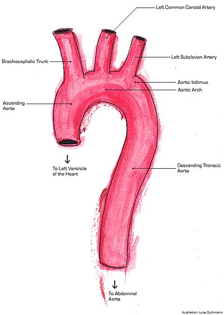
The aorta is divided into several sections: the ascending aorta, aortic arch, thoracic aorta, and abdominal aorta. Each section has distinct anatomical features and branches.

Layers of the Aorta
The aorta, like other arteries, is composed of three main layers:
- Tunica Intima: The innermost layer lined with endothelial cells.
- Tunica Media: The middle layer containing smooth muscle cells (SMCs) and elastic fibres.
- Tunica Externa (Adventitia): The outermost layer consisting of connective tissue, fibroblasts, and vasa vasorum.

Cellular Composition
The cells that make up the tissues of the aorta include:
- Endothelial Cells: Line the lumen of the aorta and form a barrier between the blood and the vessel wall.
- Smooth Muscle Cells (SMCs): Found in the tunica media, these cells provide structural support and elasticity.
- Fibroblasts: Located in the tunica externa, they secrete collagen and other extracellular matrix proteins.
- Macrophages: Present in the tunica externa, involved in immune responses and maintenance of tissue homeostasis.
Microanatomy
The aorta is classified as an elastic artery due to its high content of elastic fibres, which give it the ability to stretch and recoil. The tunica media is the thickest layer and contains concentric layers of elastic lamellae interspersed with SMCs and collagen fibres.

Histopathology
Atherosclerosis
Atherosclerosis is a common pathology of the aorta characterised by the buildup of plaques within the tunica intima. These plaques consist of lipids, inflammatory cells, SMCs, and extracellular matrix components. Over time, plaque formation can lead to vessel narrowing, reduced elasticity, and increased risk of rupture.
Aortic Aneurysm
An aortic aneurysm is a localised dilation of the aorta due to weakening of the vessel wall. Histologically, aneurysms are associated with degeneration of elastic fibres and SMCs in the tunica media, along with inflammation and fibrosis.
Aortic Dissection
Aortic dissection occurs when a tear in the tunica intima allows blood to flow between the layers of the aortic wall, creating a false lumen. Histological examination often reveals cystic medial degeneration, characterised by loss of SMCs and fragmentation of elastic fibres.
Inflammatory Conditions
Aortitis is inflammation of the aorta that can result from infections, autoimmune diseases, or trauma. Histologically, it presents with infiltration of inflammatory cells, such as lymphocytes and macrophages, into the vessel wall.
Summary of Histopathological Conditions
| Condition | Histological Features |
|---|---|
| Atherosclerosis | Plaque buildup in tunica intima, lipid-laden macrophages |
| Aortic Aneurysm | Degeneration of elastic fibres, loss of SMCs, fibrosis |
| Aortic Dissection | Tear in tunica intima, cystic medial degeneration |
| Aortitis | Inflammatory cell infiltration, vessel wall thickening |

Self-assessment MCQs (single best answer)
Which layer of the aorta is lined with endothelial cells?
What type of cells are primarily found in the tunica media of the aorta?
What is the main function of the elastic fibres in the aorta?
Which histopathological condition is characterised by plaque buildup in the tunica intima?
Which condition involves localised dilation of the aorta due to vessel wall weakening?
Which cells secrete collagen and other extracellular matrix proteins in the aorta?
What is a key histological feature of aortic dissection?
Which part of the aorta is involved in systemic circulation?
What histological changes are associated with aortic aneurysm?
Which inflammatory condition involves the infiltration of lymphocytes and macrophages into the aorta wall?
Dentaljuce
Dentaljuce provides Enhanced Continuing Professional Development (CPD) with GDC-approved Certificates for dental professionals worldwide.
Founded in 2009 by the award-winning Masters team from the School of Dentistry at the University of Birmingham, Dentaljuce has established itself as the leading platform for online CPD.
With over 100 high-quality online courses available for a single annual membership fee, Dentaljuce offers comprehensive e-learning designed for busy dental professionals.
The courses cover a complete range of topics, from clinical skills to patient communication, and are suitable for dentists, nurses, hygienists, therapists, students, and practice managers.
Dentaljuce features Dr. Aiden, a dentally trained AI-powered personal tutor available 24/7 to assist with queries and provide guidance through complex topics, enhancing the learning experience.
Check out our range of courses, or sign up now!


