Enhanced Verifiable CPD from the
University of Birmingham

Histology
Artery

Many of the slides below are optimised for colour-blind people - click the spectrum icon.

Click here to scroll down to the accompanying text and MCQs.

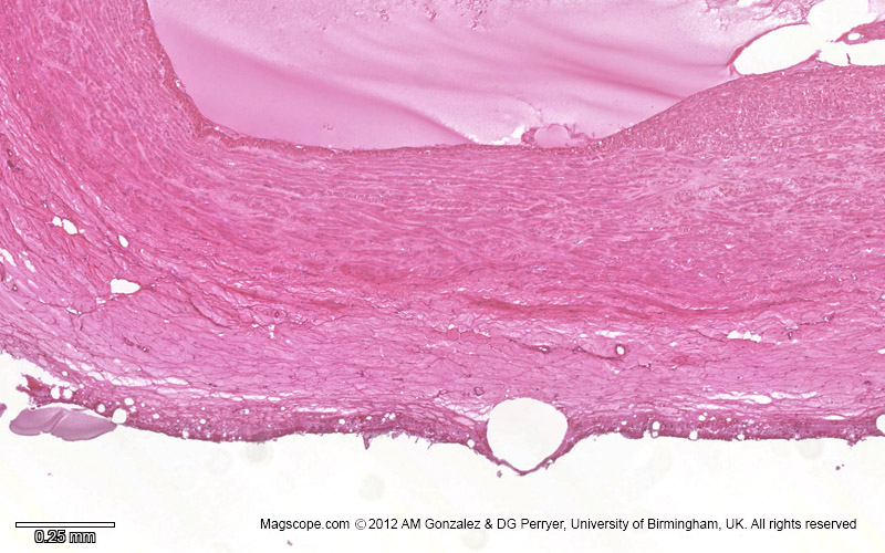
Micrograph of a cross section of an artery depicting the lumen, tunica intima, tunica media and tunica adventitia
Micrograph of a cross section of an artery illustrating the lumen, the tunica intima, tunica media and tunica adventitia
Micrograph of a cross section of an artery illustrating the lumen, the tunica intima and tunica media
Micrograph of a cross section of an artery illustrating the tunica adventitia
High magnification micrograph of a cross section of an artery illustrating the lumen, endothelium, tunica intima and tunica media
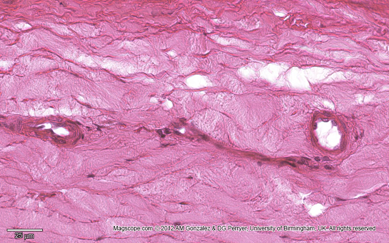
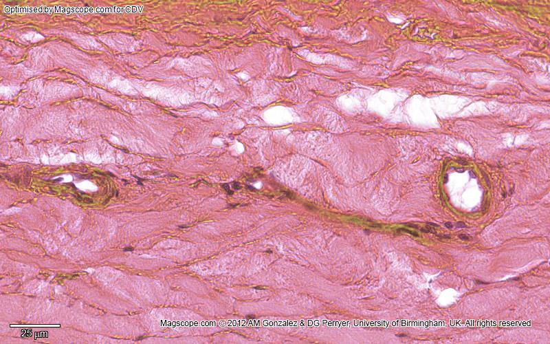
High magnification micrograph of a cross section of an artery illustrating the tunica adventitia

Artery

An artery is a blood vessel in humans and most other animals that takes oxygenated blood away from the heart in the systemic circulation to one or more parts of the body. Exceptions that carry deoxygenated blood are the pulmonary arteries in the pulmonary circulation that carry blood to the lungs for oxygenation, and the umbilical arteries in the foetal circulation that carry deoxygenated blood to the placenta. It consists of a multi-layered artery wall wrapped into a tube-shaped channel.

Arteries contrast with veins, which carry deoxygenated blood back towards the heart; or in the pulmonary and foetal circulations carry oxygenated blood to the lungs and foetus respectively.

Diagram of an artery

Structure

Microscopic anatomy of an artery.
Cross-section of a human artery.

The anatomy of arteries can be separated into gross anatomy, at the macroscopic level, and microanatomy, which must be studied with a microscope. The arterial system of the human body is divided into systemic arteries, carrying blood from the heart to the whole body, and pulmonary arteries, carrying deoxygenated blood from the heart to the lungs.

As with veins, the arterial wall consists of three layers called tunics: the tunica intima, tunica media, and tunica externa, from innermost to outermost. The tunica externa, also known as the tunica adventitia, is composed of collagen fibres and elastic tissue. The largest arteries contain vasa vasorum, small blood vessels that supply the walls of large blood vessels.

Inside this layer is the tunica media, made up of smooth muscle cells, elastic tissue (also known as connective tissue proper), and collagen fibres.

The innermost layer, the tunica intima, is in direct contact with the flow of blood and is mainly made up of endothelial cells supported by a layer of elastin-rich collagen in elastic arteries. The hollow internal cavity in which the blood flows is called the lumen.

Development

Arterial formation begins when endothelial cells start expressing arterial-specific genes, such as ephrin B2.

Function

Arteries form part of the human circulatory system.

Arteries form part of the circulatory system, carrying oxygenated blood away from the heart to the tissues. Exceptions include the pulmonary arteries, which carry blood to the lungs for oxygenation, and the umbilical arteries, which carry deoxygenated blood from a foetus to its mother. Arteries have a higher blood pressure compared to other parts of the circulatory system, which varies during the cardiac cycle, producing a pulse that can be felt in different areas of the body.

Systemic arteries can be subdivided into muscular and elastic types based on their size and the composition of their tunica media. Larger arteries are generally elastic, while smaller ones tend to be muscular. Arterial pressure is maintained by the smooth muscle contraction in the walls of arterioles, influenced by sympathetic vasomotor nerves.

Aorta

The aorta is the largest blood vessel in the human body.

The aorta is the main systemic artery, receiving blood from the left ventricle of the heart via the aortic valve. It branches into smaller arteries, arterioles, and eventually capillaries, which facilitate nutrient and gas exchange.

Capillaries

Capillaries are the smallest blood vessels, part of the microcirculation. They have a width of a single cell and lack smooth muscle, allowing for rapid diffusion of gases, sugars, and nutrients. Their small diameter provides a large surface area for exchange.

Clinical significance

Diagram showing the effects of atherosclerosis on an artery.

Systemic arterial pressures are generated by the heart's left ventricle. High blood pressure can cause arterial damage, and factors like elevated blood sugar, cholesterol, and smoking contribute to atherosclerosis. This disease is marked by the hardening of arteries due to plaque build-up. Accidental intra-arterial injection can cause severe complications, often requiring amputation.


Self-assessment MCQs (single best answer)

Which of the following blood vessels carries blood away from the heart?


Which layer of the arterial wall is in direct contact with blood flow?


What is the largest artery in the human body?


What type of blood do pulmonary arteries carry?


What is the function of capillaries in the circulatory system?


What condition is caused by the hardening of arteries due to plaque build-up?


Which type of artery is generally larger and more elastic?


What is the hollow internal cavity in an artery where blood flows called?


Which of the following can contribute to atherosclerosis?


Which arterial layer contains small blood vessels known as vasa vasorum?


Dentaljuce

Dentaljuce provides Enhanced Continuing Professional Development (CPD) with GDC-approved Certificates for dental professionals worldwide.

Founded in 2009 by the award-winning Masters team from the School of Dentistry at the University of Birmingham, Dentaljuce has established itself as the leading platform for online CPD.

With over 100 high-quality online courses available for a single annual membership fee, Dentaljuce offers comprehensive e-learning designed for busy dental professionals.

The courses cover a complete range of topics, from clinical skills to patient communication, and are suitable for dentists, nurses, hygienists, therapists, students, and practice managers.

Dentaljuce features Dr. Aiden, a dentally trained AI-powered personal tutor available 24/7 to assist with queries and provide guidance through complex topics, enhancing the learning experience.

Check out our range of courses, or sign up now!

Membership Options

Dentaljuce offers a range of membership options…

Regular Membership

With enhanced CPD Certificates. Dentaljuce is brought to you by the award winning Masters team from the School of Dentistry, University of Birmingham, UK. All have won awards for web based learning and teaching and are recognised as leaders and innovators in this field, as well as being highly experienced clinical teachers. Full access to over 150 courses, no extras to pay.

Buy Now

£89.00 per year

Student Membership

No Certificates. With universities cutting down on traditional lectures, many students are currently having to rely more on online resources. If you don't need CPD Certificates, we are offering an amazing discount on your Dentaljuce personal membership fee. Special student price just £29 for 12 months individual membership.

Buy Now

£29.00 per year

Dr. Aiden has transformed my CPD. I've learned more in the past month than I have in the last year.
MP

© Dentaljuce 2025 | Terms & Conditions | Privacy Policy

Recording CPD time: recorded.